Anatomy for first aiders: the musculoskeletal system
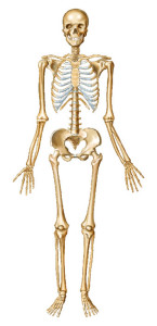
The skeletal system consists of a rigid framework of bones (206 in the adult) that perform many functions. They:
- provide structural support for the body
-
provide an anchor for muscle tendons, allowing joint movement
-
are an important site for the production of blood cells
- provide a mineral (e.g. calcium) reservoir for the body
The bones of the skeleton are connected by a series of joints. Some joints permit virtually no movement (e.g. the bones of the skull), while others are designed to permit movement (e.g. the shoulder, elbow, hip, and knee joints). Joints are held in place by fibrous bands called ligaments.
Generally, the greater the range of movement, the less stable the joint, so the shoulder joint, which allows a great range of movement, is prone to dislocation because of this.
Muscles are attached to bones via tendons. Contraction and relaxation of muscles allows movement of the bones, thereby enabling the body to move.
The skeleton consists of the:
-
skull, which encloses and protects the brain. The lower jaw, or mandible, articulates with the skull
-
spine, or vertebral column, which encloses and protects the spinal cord
-
rib cage, which protects the lungs and heart
-
bones of the upper limbs
-
pelvis
-
bones of the lower limbs
The most common injuries seen by first aiders are:
- sprains (overstretched ligaments)
- strains (overstretched muscles and tendons)
- fractures (broken bones)
- dislocations (joints out of normal position)
Want to learn how to deal with these musculoskeletal injuries? Sign up to one of our free online first aid training classes!

